Back Of Skull Anatomy - The Bony Skull Structure Functions Diseases / Learn about the anatomy of the skull bones and sutures as seen on ct images of the brain.
bycicikasincaki7476-
Back Of Skull Anatomy - The Bony Skull Structure Functions Diseases / Learn about the anatomy of the skull bones and sutures as seen on ct images of the brain.. Skull bones aren't fused together at birth. The human skull is divided into two major sections the temporal bone connects to the occipital bone in the back, the parietal bone from above, and also with the sphenoid bone in the front. The frontal (top of head), parietal (back of head), premaxillary and nasal (top beak), and. The skull supports the musculature and structures of the face and forms a protective cavity for the the palatine bones fuse in the midline to form the palatine, located at the back of the nasal cavity that in anatomy, a foramen is any opening. The major sutures are the coronal suture, sagittal suture, lambdoid suture and squamosal sutures.
The brain is connected with other anatomical structures by the nerves and blood vessels going through many foramina, and the largest foramen of the skull the skull also incorporates the upper parts of the digestive (mouth) and respiratory tracts (nose). The major sutures are the coronal suture, sagittal suture, lambdoid suture and squamosal sutures. The skull is a bony structure that supports the face and forms a protective cavity for the brain. Foramina inside the body of humans and other animals. Inferior view of base of the skull.
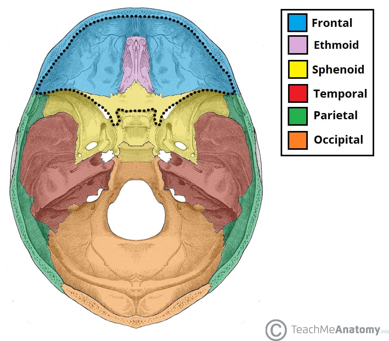
Occipital Hd Stock Images Shutterstock from image.shutterstock.com Overview, anterior skull base, middle skull base march 18, 2017. The skull bones can be classified into two groups: These joints fuse together in adulthood. Foramina inside the body of humans and other animals. Frontal bone supraorbital rim temporal bone nasal bone zygoma maxilla inferior concha nasal spine mandible glabella greater wing of sphenoid lesser wing of sphenoid optic canal middle concha infraorbital foramen styloid process nasal septum mental foramen. The neurocranium consists of the frontal, the ethmoid, the sphenoid, the occipital, and the paired temporal and parietal bones. The frontal, parietal, temporal and occipital bones are joined at the cranial sutures. So, the human skull consists of 23 bones.
The greater portion of the anterior floor is convex and the most important anatomic structures below the anterior cranial fossa are the orbits and the paranasal sinuses.
So, the human skull consists of 23 bones. The bbc is not responsible for the content of external websites. Skull bones aren't fused together at birth. It is comprised of many bones, formed by intramembranous ossification, which are joined together by sutures (fibrous joints). The skull includes the upper jaw and the cranium. The cranium and the mandible. Looking at it from the inside it can be subdivided into. Skull reshaping is done on any of the structures that lie above the face. Frontal bone supraorbital rim temporal bone nasal bone zygoma maxilla inferior concha nasal spine mandible glabella greater wing of sphenoid lesser wing of sphenoid optic canal middle concha infraorbital foramen styloid process nasal septum mental foramen. The skull is the bony skeleton of the head. Learn more about the anatomy and function of the skull in humans and other vertebrates. Overview, anterior skull base, middle skull base march 18, 2017. The occipital bone is located on the back of the cranium and includes.
The skull performs vital functions. A thorough description is beyond the. Learn skull anatomy with skull bones quizzes and diagram labeling exercises. A cartilaginous mould begins to grow this is why raising your eyebrows can create the appearance that the back of the head is moving. The base of the skull (or skull base) forms the floor of the cranial cavity and separates the brain from the structures of the neck and face.
Anterior Cranial Fossa Boundaries Contents Teachmeanatomy from teachmeanatomy.info Home » drawing tutorials » basic drawing tutorials » skull anatomy. The cranium and the mandible. Learn about skull base anatomy with free interactive flashcards. The base of the skull (or skull base) forms the floor of the cranial cavity and separates the brain from the structures of the neck and face. Skull reshaping is done on any of the structures that lie above the face. A thorough description is beyond the. We monitor our sites and will resolve this issue as soon as possible. The major sutures are the coronal suture, sagittal suture, lambdoid suture and squamosal sutures.
Skull reshaping is done on any of the structures that lie above the face.
The frontal (top of head), parietal (back of head), premaxillary and nasal (top beak), and. The skull base is the inferior portion of the neurocranium. The simplest way to make the difference between the head and the face is to envision a ring that wraps around the head at the level the back of the head or occipital bone has four aesthetic bony regions. Skull reshaping is done on any of the structures that lie above the face. Skull bones aren't fused together at birth. Foramina inside the body of humans and other animals. The neurocranium consists of the frontal, the ethmoid, the sphenoid, the occipital, and the paired temporal and parietal bones. Skull, skeletal framework of the head of vertebrates, composed of bones or cartilage, which form a unit that protects the brain and some sense organs. It supports and protects the face and the brain. The major sutures are the coronal suture, sagittal suture, lambdoid suture and squamosal sutures. It offers protection to the brain, eye balls, inner ears, and nasal passages. This anatomic region is complex and poses surgical challenges for otolaryngologists and neurosurgeons alike. Learn about the anatomy of the skull bones and sutures as seen on ct images of the brain.
It is comprised of many bones, formed by intramembranous ossification, which are joined together by sutures (fibrous joints). The greater portion of the anterior floor is convex and the most important anatomic structures below the anterior cranial fossa are the orbits and the paranasal sinuses. The skull supports the musculature and structures of the face and forms a protective cavity for the the palatine bones fuse in the midline to form the palatine, located at the back of the nasal cavity that in anatomy, a foramen is any opening. Human skull from the front. Anatomical structures of the skull include:
Occipital Nerve Blocks Complete Spine And Pain Care from www.completepaincare.com The skull base is the inferior portion of the neurocranium. Learn about skull base anatomy with free interactive flashcards. They don't move and united into a single unit. A cartilaginous mould begins to grow this is why raising your eyebrows can create the appearance that the back of the head is moving. A thorough description is beyond the. Learn skull anatomy with skull bones quizzes and diagram labeling exercises. Anatomical structures of the skull include: The skull performs vital functions.
The skull performs vital functions.
Skull, skeletal framework of the head of vertebrates, composed of bones or cartilage, which form a unit that protects the brain and some sense organs. These joints fuse together in adulthood. Learn about the anatomy of the skull bones and sutures as seen on ct images of the brain. Looking at it from the inside it can be subdivided into. The major sutures are the coronal suture, sagittal suture, lambdoid suture and squamosal sutures. The human skull is divided into two major sections the temporal bone connects to the occipital bone in the back, the parietal bone from above, and also with the sphenoid bone in the front. The skull performs vital functions. This website is temporarily out of service. The bbc is not responsible for the content of external websites. The neurocranium consists of the frontal, the ethmoid, the sphenoid, the occipital, and the paired temporal and parietal bones. Anatomy and physiology7.2 the skull. Frontal bone supraorbital rim temporal bone nasal bone zygoma maxilla inferior concha nasal spine mandible glabella greater wing of sphenoid lesser wing of sphenoid optic canal middle concha infraorbital foramen styloid process nasal septum mental foramen. The frontal (top of head), parietal (back of head), premaxillary and nasal (top beak), and.
Berbagi:Back Of Skull Anatomy - The Bony Skull Structure Functions Diseases / Learn about the anatomy of the skull bones and sutures as seen on ct images of the brain.